


Osteochondrosis is a widely distributed disease, and it is found to varying degrees in most people between the ages of 35 and 40. The most likely sites of osteonecrosis are the lower cervical spine, upper thoracic and lower lumbar spine. The etiology of osteonecrosis, that is, the specific causes and conditions of occurrence, is completely unknown. However, there are direct effects of genetic predisposition, age-related changes, trauma and impaired blood supply to tissues.
In the development of the disease, the main role is played by changes in the central part of the disc, namely dehydration. Recall that the disc consists of the medullary nucleus and the fibrous nucleus surrounding it. Due to the desiccation process, the disc loses its shock-absorbing functions, leading to gradual destruction. In the development of the disease, four stages are distinguished, differing in certain changes in the intervertebral disc and in adjacent tissues. The severity of osteonecrosis of the spine, symptoms and treatment directly depend on the stage of development of the pathology.

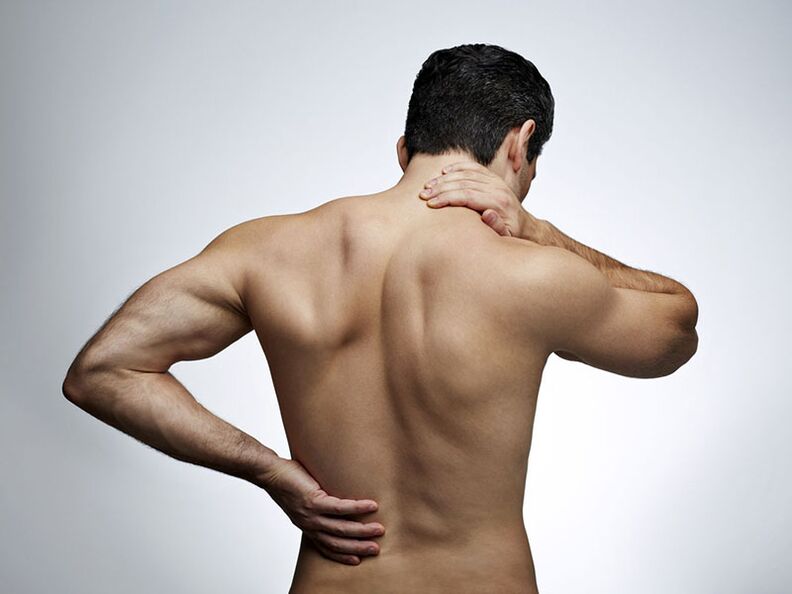
Symptoms and causes of spinal osteonecrosis
Let us briefly consider the advanced stages of osteonecrosis in terms of anatomical changes.
- First stage. The formation of cracks in the inner layers of the annulus and in the nucleus pulposus. Penetrating into the fissures, the nucleus irritates the nerve endings of the annulus;
- Second stage. While further irritation of lumbar sclerosis occurs due to pathology of the nucleus pulposus, vertebral fixation deteriorates. There is an abnormal mobility of the spine;
- Third stage. Gradual damage to annular fibrosus. There is protrusion of the nucleus (convex nucleus) beyond the anatomical boundary of the annulus, followed by annular rupture and the formation of disc herniation (extrusion);
- Fourth stage. There is a spread of degenerative changes to the surrounding tissues - vertebrae, ligaments, nerves, blood vessels. As a result of the chronic inflammatory process, the discs are scarred, which leads to the development of fibrosis.
Depending on the location in the spine, there are 3 types of osteonecrosis:
- cervical osteonecrosis;
- Thoracic osteonecrosis;
- Lumbar fibroma.
It is worth noting that several sources distinguish the fourth type - sacral necrosis.

Cervical bone necrosis |
Thoracic bone necrosis |
Lumbar bone necrosis |
|---|---|---|
|
The main types of osteonecrosis are considered cervical and lumbar. However, the thoracic spine is subjected to frequent stress and is prone to early gross degenerative changes, especially in young people. Due to the peculiarities of the development of changes in the thoracic spine, neurological symptoms manifest themselves at a late stage of the disease. In most cases, the disease occurs with an injury (eg, from heavy lifting).
|
Anomalies are commonly observed in the lumbar spine.
|
It has been mentioned before that the etiology of osteonecrosis is not completely known. However, we can name the main causes of osteonecrosis of the spine, with a proven influence on the development of the disease:
- Constant static and dynamic loads on the spine with varying intensities. For example, the work of a loader (carrying heavy objects) or a miner (in unusual positions for many hours and subject to a large amount of matter), a driver (vibrations andsedentary lifestyle) or an office worker (sedentary lifestyle);
- backache;
- Weak or overweight physical development;
- Failure to maintain correct posture and stooping;
- The result is flat feet and muscle imbalance, which leads to uneven load distribution across different parts of the spine;
- genetic traits;
- Hypothermia leads to more intense symptoms of osteonecrosis;
- And finally the human aging process.
Diagnosis of osteonecrosis of the spine
Preliminary diagnosis of the disease (osteonecrosis) occurs on the basis of the patient's complaints, examination and palpation of the spine. In addition, the affected part of the spine can be identified by the propagation topography of the pain, which can indicate the exact location (in the cervical vertebrae, thoracic, lower back) of the compression of the nerves. menstruation occurs.
The main diagnostic method for diagnosing osteonecrosis is X-ray examination. On X-ray of the spine, degenerative changes in disc joints, discs, disc stenosis, fibrous changes can be seen. stiffness in the tissues of the spine. At the same time, the recognition of spinal lesions (especially in the early stages) is always difficult, since the processes occurring in this case are characteristic of a number of other diseases (tumor, metastasis, spondylitis). ankylosing spondylitis, tuberculosis lesions).
On the anterior radiograph, irregularities of the plaques, proliferation or sharpness of uncomplicated processes are detected. In the representative image, height reduction, shape change, disc herniation, osteochondrosis and other changes in the vertebral body can be noticed. Small degenerative changes cannot be seen on plain radiographs, and contrast-enhanced X-ray examination is performed to detect them. The most complete information can be provided by discography - an x-ray examination with the introduction of contrast material through a puncture directly into the disc.
Another study of the spine is functional radiography. Thanks to X-ray films with maximum flexion and extension of the spine, it is possible to determine the mobility or immobility of the disc space.
The most modern and high-tech research methods are computed tomography (hereinafter referred to as CT) and magnetic resonance imaging (hereinafter referred to as MRI). Although the first CT and MRI machines appeared in the 70s and 80s, this technology is still being actively developed and improved, while the general population is still not accessible due to high costs or lack of access to medical equipment. machines in local hospitals.
Treatment methods for cervical spondylosis
Treatment of osteonecrosis is a complex, cyclical process. It aims to minimize and eliminate the consequences of changes in the intervertebral disc. Namely inflammation, squeezing, poor blood circulation, muscle spasms. A variety of therapies can help in this process, including those of traditional healers. In the treatment of osteonecrosis, the patient should be completely rested.
For the most part, treatment occurs by non-invasive methods, such as drug therapy, antiparasitic therapy, exercise therapy, and others. Invasive therapy, ie, surgery, is rarely used. For example, in such cases when the herniated disc persists for more than six months and conservative treatment does not give a positive effect.

Osteochondrosis of the spine and its treatment is carried out by doctors of different specialties: neurologists, orthopedists, chiropractors, orthopedists and others. However, the diagnosis, the ordering of the tests, and the studies, are usually performed by a neurologist as well as a chiropractor, if a doctor of that specialty is available in the clinic, due to the expertise of a neurologist. his about spinal diseases.
In summary, we can say that the treatment of osteonecrosis is divided into the following main categories:
- Reduce pain syndrome;
- Eliminate spasms;
- Eliminate inflammation;
- Reduce squeezing;
- Improve blood flow;
- Prevent further deterioration.
Let's take a look at commonly used treatments, each of which includes one or more of the items on this list.
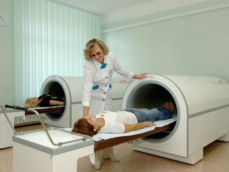
Physiotherapy treatment
Physiotherapy procedures are aimed at improving blood microcirculation in the affected spine area, eliminating pain and reducing edema. In addition, physical therapy helps reduce the dose of medication used. There are dozens of types of physical therapy. Here are some of them:
- Acupressure is impacting acupuncture points with needles, electricity, and lasers. Helps relieve spasms and improves blood flow;
- Kinesitherapy is a treatment through movement and special physical exercises. This is, in fact, gymnastics, but in an extended sense. This includes simulation training, group exercises under the guidance of a physician, spinal stretching;
- Therapeutic swimming. Allows you to perform exercises that are harmful to the spine for patients who find it difficult to do them. For example, the elderly, people who are overweight or have mobility disabilities. It is important to note that due to the lifting force of the water, there is an even distribution of the load on the spine;
- Electrotherapy, UHF therapy, nerve stimulation, magnetic therapy, massage, and others.
Acupuncture
A unique method from oriental medicine. Its essence lies in the use of special very thin needles that enter particular points of the body, literally, under the influence of gravity. These points were discovered by the thousands of years of experience of Eastern healers, later proven by orthodox medicine. Acupuncture eliminates even severe pain, has a positive effect on the general nervous system and neurological diseases.
Surgical intervention
Surgery is indicated only if there is no positive effect from non-invasive treatment or in case of severe complications. The main surgical treatment is discectomy - removing the damaged disc. However, even after minimally invasive surgery, rehabilitation will take at least 6 months.
At the same time, surgery will not eliminate the need for regular treatment of osteonecrosis. Since before surgery, there is always the obvious task of eliminating a specific defect: hernia, deformity, hernia, etc. v. The activity does not affect the general process of degeneration of the cartilage tissue of the spine.
Medical treatment
This is the most effective type of treatment in the short term. Instant pain reliever, anti-inflammatory, spasm reliever, etc. v. However, as soon as they are canceled, in the absence of other procedures needed to treat osteonecrosis, their effects quickly wear off. At the same time, you can not spend your life on drugs, sooner or later they will cause side effects on the body.

Medicines can be classified as primary and secondary treatment. They are often needed to improve patients' lives. They are used to relieve pain (therapeutic blockade), eliminate inflammation, relieve muscle spasms, improve blood flow. In recent years, another drug is increasingly used - chondroprotectors. However, the drug alone cannot achieve a permanent lasting effect.
Treatment at home
Home treatment of osteonecrosis includes manual therapy (exercise therapy, collar of Shants), acupuncture, and vacuum therapy. These methods help improve blood circulation and relieve congestion in the muscles and spine. As a result, tissue regeneration is significantly improved.
The listed procedures in combination with medication provide an effective treatment that relieves unpleasant symptoms and improves the general condition. In this case, special attention should be paid to proper nutrition and regulation of excess weight.
Physical therapy
Thanks to exercise, mobility of the vertebrae is restored, their muscles and circulatory system are strengthened. The latter is of great importance, because the spine is deprived of its blood supply system and the discs can only receive nutrients through nearby tissues. In this case, it is necessary to distinguish between therapeutic exercises and any other way of training.

Manual therapy
One of the most effective methods. Because manual therapists work on muscles, ligaments, and bones. It improves posture, restores the "normal" structure of the skeleton, relieves excess muscle tension. Including, it directly affects the vertebrae.
Manual osteolysis should be performed on a regular basis, from once a year to several times, depending on the need. It should be noted that this is not a one-time, multi-way, 10-20 session course. Only then will it have a lasting positive effect.